
Rubella Virus Antigens
Rubella virus (RV) also known as German measles, is the only member of the genus Rubivirus and belongs to the family of Togaviridae. The greatest threat of rubella virus to public health is its teratogenicity. Pregnant women who infected with RV can cause congenital harm on infants, especially to the developing baby in the first 3 months of pregnancy. RV is spread through blood to fetus, resulting in spontaneous abortion, stillbirth and fetal infection. Congenital rubella syndrome (CRS) is an important cause of blindness, deafness, congenital heart disease and mental retardation. It is estimated that more than 100,000 infants are born with CRS each year worldwide. RV mostly affects children, adolescents and young adults. RV can also cause transient joint involvement such as polyarthralgia and arthritis.
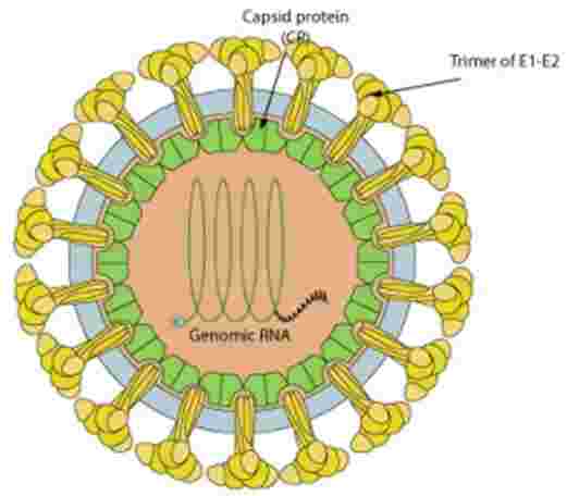
Rubella virus is a positive-strand RNA virus. Its genome consists of a single-stranded 40 S RNA of positive polarity. In addition to the 40 S genomic RNA, RV-infected cells contain a subgenomic 24 S RNA derived from the 3' end of the 40 S RNA. The translation of the 24 S subgenomic RNA produces a 110-kDa precusor polyprotein that is proteolytically processed to yield three structural proteins, capsid protein C, type 1 membrane glycoproteins E2 and E1. E1 (58 kDa) and E2 (42-47 kDa) are both found on the virion surface as viral spikes. Studies with monoclonal antibodies (MAbs) suggest that E1 is the major target for antibodies. E2 is topologically buried under E1 on the viral surface and plays a role in the cell surface expression of E1.
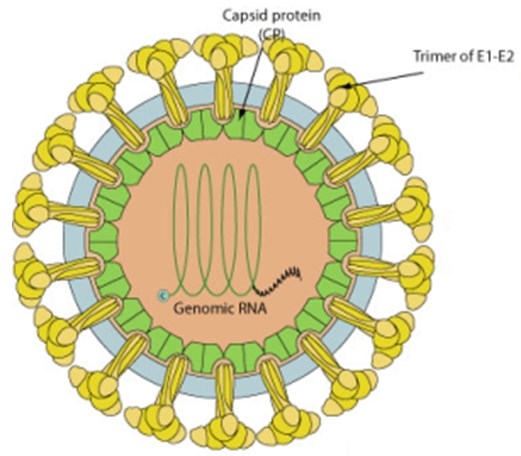
Fig 1. The structure of Rubella Virus (Wang-Shick Ryu. 2017)
Rubella virus particles enter target cells via clathrin-mediated endocytosis (Figure 2). Fusion of the viral and endosome membranes occurs in a low pH- and calcium-dependent reaction and is mediated by the E1 membrane fusion protein. Viral RNA replication occurs in the cytoplasm. The nascent NC, E2, and E1 assemble on and bud into the Golgi complex to form progeny virions, which are transported through the secretory pathway and released at the plasma membrane.
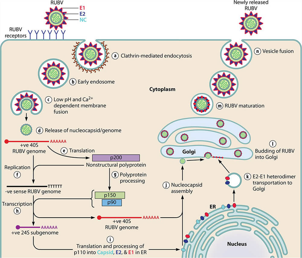
Fig 2. Model of the Rubella Virus life cycle1
Creative Diagnostics is one of the best manufactures of viral antigens, customers from all over the world find their satisfactory with our product and service. Welcome to contact our sales representative if you are in need of rubella virus antigens: Rubella capsid proteins, Rubella E1, E2 proteins, RuV Envelope Protein 1, RuV E1 Mosaic proteins and RuV Active E1/E2 proteins.
References
- Wang-Shick Ryu. (2017). Molecular Virology of Human Pathogenic Viruses.
- Das, P.K.; Kielian, M. (2021) Molecular and Structural Insights into the Life Cycle of Rubella Virus. Journal of Virology, 95(10), e02349-20